Tag Archive for: Biology
 https://www.saffronwaldenmuseum.org/wp-content/uploads/2023/02/Saffron_Walden_Museum_deer-calcaneus.jpg
624
832
swmuseum
https://www.saffronwaldenmuseum.org/wp-content/uploads/2023/02/Saffron-Walden-Museum-Logo-.png
swmuseum2023-02-14 14:59:322023-07-07 10:33:04Identification – deer calcaneus
https://www.saffronwaldenmuseum.org/wp-content/uploads/2023/02/Saffron_Walden_Museum_deer-calcaneus.jpg
624
832
swmuseum
https://www.saffronwaldenmuseum.org/wp-content/uploads/2023/02/Saffron-Walden-Museum-Logo-.png
swmuseum2023-02-14 14:59:322023-07-07 10:33:04Identification – deer calcaneus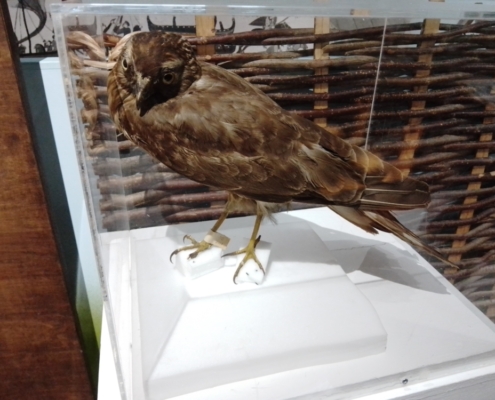 https://www.saffronwaldenmuseum.org/wp-content/uploads/2023/03/Hen-harrier-OoM-display-scaled-e1685959467497.jpg
750
1000
swmuseum
https://www.saffronwaldenmuseum.org/wp-content/uploads/2023/02/Saffron-Walden-Museum-Logo-.png
swmuseum2022-06-10 08:58:452023-07-07 10:33:08Object of the Month – June 2022
https://www.saffronwaldenmuseum.org/wp-content/uploads/2023/03/Hen-harrier-OoM-display-scaled-e1685959467497.jpg
750
1000
swmuseum
https://www.saffronwaldenmuseum.org/wp-content/uploads/2023/02/Saffron-Walden-Museum-Logo-.png
swmuseum2022-06-10 08:58:452023-07-07 10:33:08Object of the Month – June 2022 https://www.saffronwaldenmuseum.org/wp-content/uploads/2023/03/IMG_20211001_121435-scaled.jpg
1920
2560
swmuseum
https://www.saffronwaldenmuseum.org/wp-content/uploads/2023/02/Saffron-Walden-Museum-Logo-.png
swmuseum2021-10-01 13:46:512023-07-13 11:34:42Object of the Month – October 2021
https://www.saffronwaldenmuseum.org/wp-content/uploads/2023/03/IMG_20211001_121435-scaled.jpg
1920
2560
swmuseum
https://www.saffronwaldenmuseum.org/wp-content/uploads/2023/02/Saffron-Walden-Museum-Logo-.png
swmuseum2021-10-01 13:46:512023-07-13 11:34:42Object of the Month – October 2021 https://www.saffronwaldenmuseum.org/wp-content/uploads/2018/08/saffron-walden-museum-A-Red-squirrel.jpeg
476
624
swmuseum
https://www.saffronwaldenmuseum.org/wp-content/uploads/2023/02/Saffron-Walden-Museum-Logo-.png
swmuseum2018-08-01 10:00:542023-07-12 11:49:27Object of the Month – August 2018
https://www.saffronwaldenmuseum.org/wp-content/uploads/2018/08/saffron-walden-museum-A-Red-squirrel.jpeg
476
624
swmuseum
https://www.saffronwaldenmuseum.org/wp-content/uploads/2023/02/Saffron-Walden-Museum-Logo-.png
swmuseum2018-08-01 10:00:542023-07-12 11:49:27Object of the Month – August 2018