Tag Archive for: British wildlife
 https://www.saffronwaldenmuseum.org/wp-content/uploads/2023/06/saffron-walden-museum-dragonfly-larva-skin.jpeg
768
1024
Jess
https://www.saffronwaldenmuseum.org/wp-content/uploads/2023/02/Saffron-Walden-Museum-Logo-.png
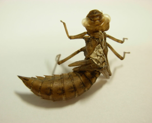
Jess2023-06-09 15:03:082023-07-07 10:33:00Object of the Month – June 2023
https://www.saffronwaldenmuseum.org/wp-content/uploads/2023/06/saffron-walden-museum-dragonfly-larva-skin.jpeg
768
1024
Jess
https://www.saffronwaldenmuseum.org/wp-content/uploads/2023/02/Saffron-Walden-Museum-Logo-.png
Jess2023-06-09 15:03:082023-07-07 10:33:00Object of the Month – June 2023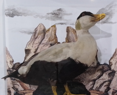 https://www.saffronwaldenmuseum.org/wp-content/uploads/2023/03/20-Common-eider-img-cropped-e1685959004835.jpg
1010
1000
swmuseum
https://www.saffronwaldenmuseum.org/wp-content/uploads/2023/02/Saffron-Walden-Museum-Logo-.png
swmuseum2023-01-31 11:29:472023-07-12 14:08:17Object of the Month – February 2023
https://www.saffronwaldenmuseum.org/wp-content/uploads/2023/03/20-Common-eider-img-cropped-e1685959004835.jpg
1010
1000
swmuseum
https://www.saffronwaldenmuseum.org/wp-content/uploads/2023/02/Saffron-Walden-Museum-Logo-.png
swmuseum2023-01-31 11:29:472023-07-12 14:08:17Object of the Month – February 2023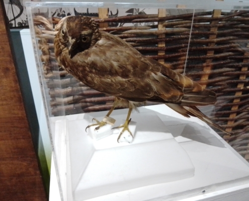 https://www.saffronwaldenmuseum.org/wp-content/uploads/2023/03/Hen-harrier-OoM-display-scaled-e1685959467497.jpg
750
1000
swmuseum
https://www.saffronwaldenmuseum.org/wp-content/uploads/2023/02/Saffron-Walden-Museum-Logo-.png
swmuseum2022-06-10 08:58:452023-07-07 10:33:08Object of the Month – June 2022
https://www.saffronwaldenmuseum.org/wp-content/uploads/2023/03/Hen-harrier-OoM-display-scaled-e1685959467497.jpg
750
1000
swmuseum
https://www.saffronwaldenmuseum.org/wp-content/uploads/2023/02/Saffron-Walden-Museum-Logo-.png
swmuseum2022-06-10 08:58:452023-07-07 10:33:08Object of the Month – June 2022 https://www.saffronwaldenmuseum.org/wp-content/uploads/2023/03/Water-vole-c-Safforn-Walden-Museum.jpg
1681
2557
swmuseum
https://www.saffronwaldenmuseum.org/wp-content/uploads/2023/02/Saffron-Walden-Museum-Logo-.png
swmuseum2021-02-05 16:35:102023-07-07 10:33:14Object of the Month – February 2021
https://www.saffronwaldenmuseum.org/wp-content/uploads/2023/03/Water-vole-c-Safforn-Walden-Museum.jpg
1681
2557
swmuseum
https://www.saffronwaldenmuseum.org/wp-content/uploads/2023/02/Saffron-Walden-Museum-Logo-.png
swmuseum2021-02-05 16:35:102023-07-07 10:33:14Object of the Month – February 2021 https://www.saffronwaldenmuseum.org/wp-content/uploads/2023/03/Promicroceras_pyritosum-Ammojoe-CC-BY-SA-https-creativecommons.org-licenses-by-sa-3.0-scaled-e1685613885342.jpg
750
1000
swmuseum
https://www.saffronwaldenmuseum.org/wp-content/uploads/2023/02/Saffron-Walden-Museum-Logo-.png
swmuseum2020-02-07 11:57:372023-07-11 14:47:44Identification – Ammonite in sandstone
https://www.saffronwaldenmuseum.org/wp-content/uploads/2023/03/Promicroceras_pyritosum-Ammojoe-CC-BY-SA-https-creativecommons.org-licenses-by-sa-3.0-scaled-e1685613885342.jpg
750
1000
swmuseum
https://www.saffronwaldenmuseum.org/wp-content/uploads/2023/02/Saffron-Walden-Museum-Logo-.png
swmuseum2020-02-07 11:57:372023-07-11 14:47:44Identification – Ammonite in sandstone https://www.saffronwaldenmuseum.org/wp-content/uploads/2023/03/0-Main-Image-v2-e1685613792109.jpg
749
1000
swmuseum
https://www.saffronwaldenmuseum.org/wp-content/uploads/2023/02/Saffron-Walden-Museum-Logo-.png
swmuseum2019-10-07 12:06:362023-07-07 10:33:21Object of the Month – October 2019
https://www.saffronwaldenmuseum.org/wp-content/uploads/2023/03/0-Main-Image-v2-e1685613792109.jpg
749
1000
swmuseum
https://www.saffronwaldenmuseum.org/wp-content/uploads/2023/02/Saffron-Walden-Museum-Logo-.png
swmuseum2019-10-07 12:06:362023-07-07 10:33:21Object of the Month – October 2019 https://www.saffronwaldenmuseum.org/wp-content/uploads/2023/03/Cabbage-whites-SAFWM-118007-crop-e1685613728615.jpg
1262
1000
swmuseum
https://www.saffronwaldenmuseum.org/wp-content/uploads/2023/02/Saffron-Walden-Museum-Logo-.png
swmuseum2019-06-22 15:08:192023-07-12 14:52:32Object of the Month – June 2019
https://www.saffronwaldenmuseum.org/wp-content/uploads/2023/03/Cabbage-whites-SAFWM-118007-crop-e1685613728615.jpg
1262
1000
swmuseum
https://www.saffronwaldenmuseum.org/wp-content/uploads/2023/02/Saffron-Walden-Museum-Logo-.png
swmuseum2019-06-22 15:08:192023-07-12 14:52:32Object of the Month – June 2019